Principales conclusiones
- The portable ultrasound market is valued at USD 2.49 billion in 2025 and projected to reach USD 3.84 billion by 2030 at 9.0% CAGR — edge AI hardware is the primary enabler (MarketsandMarkets)
- The AI-in-ultrasound market is growing at 24% CAGR, reaching USD 6.88 billion by 2030 — driven by automated diagnostics and point-of-care deployment
- RK3588's 48MP ISP 3.0 handles ultrasound probe signal preprocessing, HDR imaging, and multi-camera capture simultaneously — without external image processing hardware
- La 6 TOPS NPU funciona diagnostic AI models (nodule detection, measurement automation, anomaly scoring) at real-time speeds on-device — eliminating cloud dependency for offline clinical deployments
- 8K H.265 hardware encoding generates DICOM-compatible image archives at high compression ratios — reducing storage requirements by 40–60% vs. uncompressed DICOM
- RK3588-based medical device designs achieve IEC 60601-1 electrical safety compliance through proper isolation design — the SoC itself is not the certification bottleneck
Why Medical Imaging Is Moving to Edge-Embedded AI Platforms
Medical imaging is undergoing a structural transformation. According to MarketsandMarkets, the global portable ultrasound market was valued at USD 2.49 billion in 2025 and is projected to reach USD 3.84 billion by 2030, driven by decentralized care delivery, an aging global population, and the compelling economics of point-of-care diagnostics.
The hospital radiology suite is no longer the only setting in which high-quality ultrasound diagnostics can be performed. Emergency departments, rural clinics, home healthcare visits, and remote community health programs are all emerging as deployment environments — each demanding a device profile that traditional cart-based imaging systems cannot satisfy: compact, battery-powered, and capable of AI-assisted interpretation without cloud connectivity.
The Role of Embedded AI in Modern Medical Devices
AI integration in medical imaging is no longer a premium feature reserved for flagship hospital systems. Research from SNS Insider reports that 88% of clinical settings worldwide have integrated AI-powered ultrasound solutions, with deep learning algorithms automating measurements, reducing operator dependency, and improving diagnostic accuracy in cardiology, obstetrics, and emergency medicine.
For medical device manufacturers, this market evolution creates a specific engineering challenge: how to embed AI inference capable of clinical-grade diagnostic support into a device that is portable, battery-operated, thermally sealed, and manufacturable at a cost point appropriate for mid-market and emerging market healthcare deployment. The RK3588 addresses this challenge with a hardware architecture that is unusually well-matched to the requirements of portable medical imaging.
Why Legacy Embedded Platforms Are Insufficient
Previous-generation ARM SoCs — Cortex-A72-based platforms, i.MX8M Plus, and similar mid-range embedded processors — provide adequate performance for basic medical device HMI and data acquisition. They are insufficient for real-time AI-assisted diagnostics. Without a dedicated NPU, running a diagnostic assistance model alongside image acquisition and display rendering forces a choice between acceptable frame rates and acceptable inference speed. The RK3588 eliminates this trade-off with parallel CPU, GPU, NPU, and ISP execution.
RK3588 Hardware Architecture for Medical Imaging Applications
Four specific hardware subsystems within the RK3588 SoC are directly relevant to medical imaging device design. Understanding each subsystem's medical device application prevents over-engineering in some areas and under-specifying in others.
ISP 3.0: The 48MP Image Signal Processor
The RK3588 integrates a dual-pipe ISP 3.0 supporting sensors up to 48MP. In medical imaging, the ISP's role extends beyond simple image capture. Ultrasound probe outputs require signal conditioning, noise filtering, and dynamic range compression before AI processing. The dual-pipe architecture supports simultaneous processing of two independent camera streams — for example, a primary diagnostic ultrasound view alongside a secondary anatomical reference camera, both processed without CPU involvement.
NPU: 6 TOPS for Clinical AI Inference
The 6 TOPS NPU is the defining capability for AI-assisted medical imaging on the RK3588. Diagnostic assistance models — nodule detection classifiers, automated measurement tools, anomaly scoring networks — are typically deployed as INT8 quantized models in the 5–50MB weight range. Clinical deployment advantages of on-device NPU inference include: no cloud connectivity requirement, deterministic latency regardless of network conditions, and elimination of patient data transmission to third-party servers — a significant consideration under GDPR and HIPAA. For detailed NPU benchmarks, see our Guía de rendimiento de la NPU RK3588.
Video Codec: 8K H.265 Hardware Encoding for DICOM
DICOM supports H.264 and H.265 video compression for dynamic imaging modalities. The RK3588's hardware H.265 encoder — supporting 8K@30fps — generates DICOM-compliant video archives with 40–60% smaller file sizes than equivalent H.264 encoding, directly reducing storage costs. Hardware encoding offloads this entirely from the CPU, leaving it free for AI inference and UI rendering.
Display and Touch Interface Capabilities
The RK3588 supports up to four simultaneous display outputs at resolutions up to 4K, with eDP for embedded panel integration, MIPI-DSI for compact displays, and HDMI 2.1 for external monitors. Mali-G610 GPU handles Qt-based clinical interfaces, real-time image overlays, and measurement annotation tools without CPU involvement.
| Medical Device Subsystem | RK3588 Hardware | Especificación | Clinical Benefit |
|---|---|---|---|
| Probe signal processing | ISP 3.0 dual-pipe | 48MP, HDR, multi-camera | Hardware preprocessing, no CPU load |
| AI diagnostic assistance | 6 TOPS NPU | INT4/INT8/FP16, RKNN-Toolkit2 | On-device inference, no cloud needed |
| Image archive (DICOM) | H.265 HW encoder | 8K@30fps, 4K@120fps | 40–60% smaller DICOM files |
| Clinical display | Mali-G610 GPU | 4× outputs, 4K, eDP/MIPI/HDMI | Smooth UI + real-time annotation |
| Data connectivity | PCIe 3.0 + 2×GbE + USB 3.1 | Multi-interface | PACS integration, wireless module |
| Power management | 8nm process, DVFS | 5–13W full load | Battery-powered portable devices |
| Storage (patient data) | eMMC 5.1 + NVMe via PCIe | Up to 256GB eMMC | Local DICOM archive, no external drive |
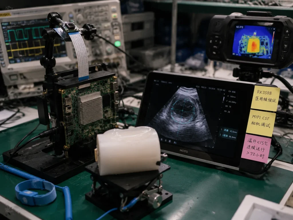
Supported Medical Imaging Applications on RK3588
The RK3588's hardware profile maps to a specific set of medical imaging device categories where its capabilities are fully utilized. Understanding this mapping prevents under-specifying (sacrificing AI capability) or over-specifying (creating an unsolvable thermal management problem in a portable enclosure).
Point-of-Care Ultrasound (POCUS) Terminals
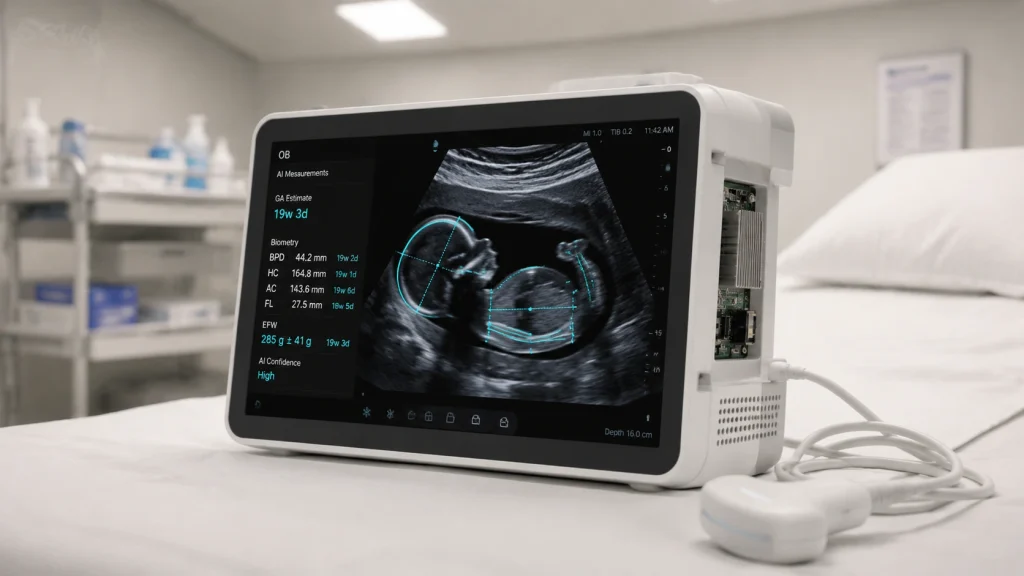
POCUS devices are used in emergency medicine, obstetrics, and anesthesiology. They require real-time ultrasound image rendering at 30–60fps, AI-assisted measurement automation (fetal biometry, ejection fraction), and local DICOM storage with optional WiFi transmission to PACS. The RK3588 handles all workloads simultaneously — ISP processes probe data, NPU runs AI models, GPU renders the clinical display — with total system power under 10W enabling 4–8 hours of battery operation.
Companies such as Butterfly Network y Fujifilm Sonosite have pioneered handheld POCUS, demonstrating clinical acceptance of portable AI-integrated ultrasound — a market now accessible to OEM/ODM manufacturers building on platforms like the RK3588.
AI-Assisted Diagnostic Workstations
Bedside diagnostic workstations for ICU, ward, and outpatient settings require larger displays (15–21"), higher compute for complex AI models, and redundant hospital network connectivity. The RK3588 serves this segment through its 4K display output, 32GB LPDDR4X maximum RAM, PCIe 3.0 for optional AI accelerator expansion, and dual Gigabit Ethernet.
Portable Endoscopy Systems
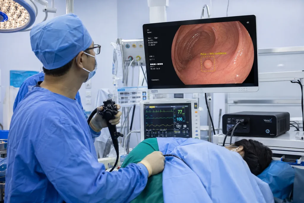
The RK3588's MIPI CSI-2 interface connects directly to endoscopic camera modules at 4K resolution. The NPU runs polyp detection models (YOLOv8 or custom CNNs at 30+ FPS), and the H.265 hardware encoder archives the procedure in DICOM-compliant format. Total system power under 15W enables battery-powered portable colonoscopy units for field screening programs.
Bedside Patient Monitoring Systems
Multi-parameter patient monitors aggregate ECG, SpO2, blood pressure, temperature, and respiration data. The RK3588's multiple UART and I2C interfaces handle sensor acquisition, the NPU runs arrhythmia classification models, and the GPU renders real-time waveform displays — making it a capable platform for next-generation AI-integrated bedside monitoring.
DICOM Integration and PACS Connectivity on RK3588
DICOM compliance is non-negotiable for medical imaging devices deployed in clinical settings. The RK3588 runs Debian 12 or Ubuntu 22.04, both of which support the mature open-source DCMTK (DICOM Toolkit) — developed by OFFIS and the Regensburg University Hospital — providing DICOM file creation, C-STORE/C-FIND/C-MOVE network operations, and DICOM conformance statement generation.
# Install DCMTK on RK3588 Debian 12
sudo apt install dcmtk libdcmtk-dev
# Convert captured image to DICOM format
img2dcm -i JPEG input_image.jpg output.dcm
# Send DICOM file to hospital PACS via C-STORE
storescu -aec PACS_AET 192.168.1.100 104 output.dcm
# Query PACS for existing studies (C-FIND)
findscu -S -k QueryRetrieveLevel=STUDY \
-k PatientID="12345" \
192.168.1.100 104
PACS Connectivity Architecture
The standard workflow: image acquisition on device → local DICOM file creation via DCMTK → C-STORE transmission to PACS server → PACS acknowledgment → local cache management. The RK3588's dual Gigabit Ethernet supports both hospital LAN and isolated device management network simultaneously, with WiFi 6 available for wireless PACS connectivity.
HL7 FHIR Integration for Electronic Health Records
Modern hospital workflows increasingly require devices to exchange data with EHR systems via HL7 FHIR APIs. The RK3588 running Linux supports HAPI FHIR (Java) and py-fhirclient (Python), enabling devices to automatically associate images with patient records and trigger clinical workflow events on image capture.
Regulatory Considerations: IEC 60601, FDA, and CE MDR
The RK3588 is an embedded computing component, not a medical device — regulatory obligations apply to the finished product. Understanding this distinction prevents both unnecessary alarm and false reassurance.
Electrical Safety
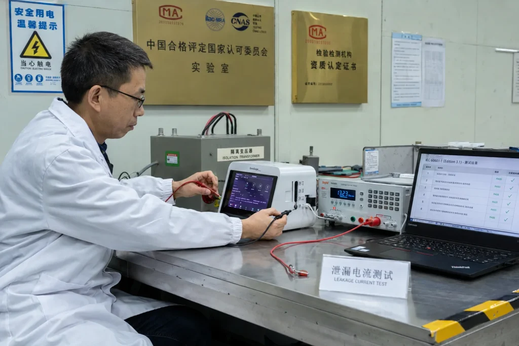
General safety requirements for medical electrical equipment. Governs isolation, leakage current, and creepage distances between patient-connected circuits and mains power.
Software Lifecycle
Medical device software development lifecycle requirements. Applies to AI diagnostic software running on the RK3588 — not to the embedded Linux OS or BSP unless classified as medical software.
US Market Clearance
Predicate-based clearance pathway for Class II medical devices. AI-assisted diagnostic functions may require De Novo classification or PMA depending on intended use.
EU Market Access
EU Medical Device Regulation requires conformity assessment by a Notified Body for Class IIa and above devices. RK3588-based imaging devices typically fall under Class IIa or IIb.
Quality Management
Quality management system standard for medical device manufacturers. Required for CE marking and FDA registration. Applies to the device manufacturer's processes, not component suppliers.
Data Privacy
On-device NPU inference (no cloud transmission) simplifies HIPAA/GDPR compliance significantly — no protected health information leaves the device during AI processing.
Important: Board vs. Device Certification
The RK3588 development board or SoM is not IEC 60601-certified and is not a medical device. Regulatory certification applies to the finished product including enclosure, power supply, patient-connected circuits, and software. ieeker can provide component-level compliance documentation (CE, FCC, RoHS) to support the device-level certification process.
Practical Isolation Architecture for IEC 60601-1 Compliance
The standard approach: isolated power supply for patient-connected front-end circuits, optical isolation or digital isolators on signal lines crossing the isolation barrier, and galvanic isolation on USB or serial interfaces connected to patient-facing peripherals. The RK3588 SoC operates entirely on the non-patient side of the isolation barrier in a properly designed carrier board.
Deploying AI Diagnostic Models on RK3588: Clinical Inference Pipeline
The pathway from a trained diagnostic AI model to a production medical imaging device involves three engineering stages, each with medical-device-specific considerations that differ from standard industrial AI deployment.
Stage 1: Model Training and Validation (Off-Device)
Diagnostic AI models are trained on clinical datasets using PyTorch or TensorFlow. Model architecture selection follows different criteria than industrial vision: sensitivity and specificity metrics are primary (not just mAP), false negative rates are often weighted more heavily than false positives, and model interpretability (Grad-CAM visualizations) is frequently required for regulatory submissions. Common architectures include EfficientNet for classification, U-Net for segmentation, and lightweight DETR variants for detection.
Stage 2: Quantization and RKNN Conversion
Converting a medical AI model to RKNN format follows the RKNN-Toolkit2 workflow, with one critical addition: post-quantization clinical validation. An INT8 quantization that reduces accuracy by 0.5% is acceptable in machine vision inspection. In a medical diagnostic context, a 0.5% drop on a nodule detection model may have patient safety implications and must be validated against the same clinical metrics used in original training.
from rknn.api import RKNN
rknn = RKNN(verbose=True)
rknn.load_onnx(model='./thyroid_nodule_detector.onnx')
rknn.config(
mean_values=[[123.675, 116.28, 103.53]],
std_values=[[58.395, 57.12, 57.375]],
target_platform='rk3588',
# Use clinical validation set — minimum 500 annotated cases
# covering full range of patient demographics and imaging conditions
)
rknn.build(
do_quantization=True,
dataset='./clinical_calibration_dataset.txt'
)
# Validate sensitivity/specificity on held-out clinical test set
# before production deployment — not just mAP or accuracy
rknn.export_rknn('./thyroid_nodule_detector_int8.rknn')
Stage 3: Clinical Inference Integration
On the RK3588 device, the AI inference pipeline runs as a background service with real-time output fed to the clinical display. The NPU inference thread operates at SCHED_FIFO priority to ensure consistent latency — a clinician should never experience inference result delays caused by background OS activity during an active diagnostic session.
Solving an ISP Calibration Problem on a Portable Ultrasound Terminal
A medical device OEM developing a portable point-of-care ultrasound terminal contacted us eight weeks before their clinical validation submission deadline with a display quality problem. Their device — targeting obstetrics and emergency medicine in Southeast Asian clinic networks — used an RK3588 SBC as the computing core, with a digitized ultrasound probe signal fed into the ISP via a custom analog front-end board.
The problem emerged during clinical evaluation: radiologists reported inconsistent brightness and contrast between devices of the same model. Two units from the same production batch produced visually distinct images from the same probe on the same patient. Signal analysis ruled out probe variance — the issue was occurring in the ISP processing stage.
Diagnosis revealed that the RK3588 ISP's Auto White Balance parameters had been left at factory defaults, which assume a standard visible-light photographic scene. Ultrasound image data has a fundamentally different spectral distribution — the default AWB algorithm introduced systematic bias in speckle-heavy ultrasound images, amplified by minor manufacturing tolerances in the analog front-end components.
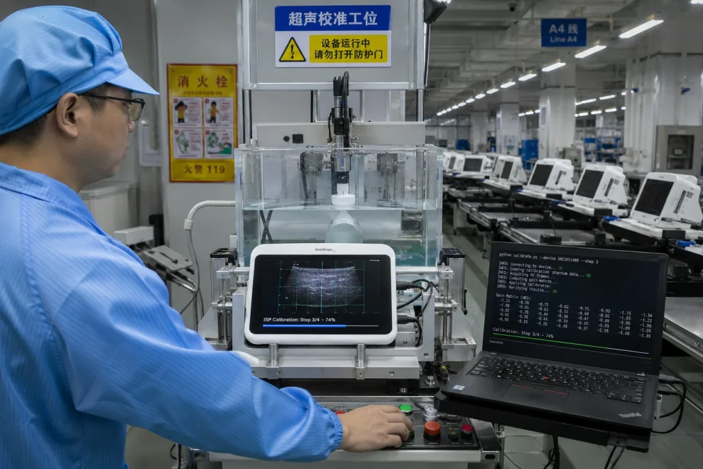
The fix required custom ISP tuning: disabling automatic AWB for ultrasound mode, calibrating a fixed gain matrix per device unit using a standard test phantom during production, and applying calibrated parameters as device-specific ISP configuration at boot. We developed a 20-second automated calibration fixture run on each unit at end of line.
The lesson for medical device teams: the ISP requires application-specific tuning for non-photographic imaging modalities. Factor ISP calibration into your production process from the beginning — retrofitting it late in development is expensive.
50-Unit Portable Ultrasound Terminal Deployment for Rural Primary Care
In Q2 2024, a digital health NGO partnering with a regional government health program contracted a medical device OEM to supply 50 portable ultrasound diagnostic terminals for rural East Africa primary care clinics. The deployment context was demanding: no reliable internet connectivity, unreliable power grid (requiring 6+ hours battery operation), ambient temperatures up to 40°C, and clinical staff with limited ultrasound training who needed AI assistance to compensate for the absence of trained sonographers.
The device used an ieeker RK3588 industrial SBC. The AI inference stack ran three concurrent models on the NPU: a fetal biometry automation model, an obstetric complication screening model, and an image quality assessment model that guided untrained operators to achieve diagnostic-quality probe positioning.
The operator guidance model — evaluating ultrasound image quality in real time and displaying directional prompts — was the most clinically impactful feature. Staff with one hour of training consistently achieved diagnostic-quality images within 3–5 minutes per examination, compared to 15–25 minutes for equivalent manual guidance by experienced sonographers.
The 8.9W full-system power draw was the enabling constraint for the battery specification. At 50–80W for a typical x86 alternative, the same battery pack would last under 2 hours. The RK3588's power profile was not a convenience feature — it was the physical enabler of the product's clinical utility.
RK3588 vs. Alternative Platforms for Medical Imaging
| Plataforma | AI Compute | ISP | Potencia | Medical Device Fit |
|---|---|---|---|---|
| RK3588 | 6 TOPS NPU | Dual 48MP ISP 3.0 | 5-13W | POCUS · Endoscopy · Bedside Monitor |
| i.MX8M Plus (NXP) | 2.3 TOPS NPU | Dual ISP (limited HDR) | 3–6W | Patient Monitor · Low-complexity AI |
| Jetson Orin Nano | 40 TOPS GPU | No ISP | 7-15W | High-complexity AI · Needs ext. ISP |
| Snapdragon 888 | 26 TOPS (Hexagon) | Triple ISP (Spectra 580) | 5–12W | Handheld · Medical BSP limited |
| x86 (Core i5/i7) | None (dGPU add-on) | None (ext. frame grabber) | 35–80W | Cart systems only · Not portable |
For applications requiring real-time AI diagnostic assistance alongside high-resolution imaging, the RK3588's 6 TOPS NPU and 48MP ISP combination is the most capable option in the sub-15W power envelope currently available in production-ready industrial SBC form factors.
Board Form Factors for Medical Device Integration
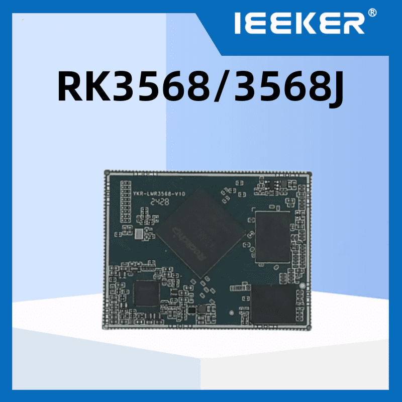
Medical device hardware engineers face a form factor decision with additional constraints: IEC 60601-1 isolation requirements favor keeping patient-connected and non-patient circuits on separate boards, and regulatory documentation benefits from using a commercial SoM as a clearly defined off-the-shelf component with its own CE and FCC declarations.
🧩 SoM + Custom Medical Carrier Board
- Cleanest regulatory boundary — SoM is OTS component
- Custom isolation design on carrier board
- Optimized form factor for device enclosure
- Ideal for Class IIa/IIb devices (510k / CE MDR)
- Recommended for volume >200 units/year
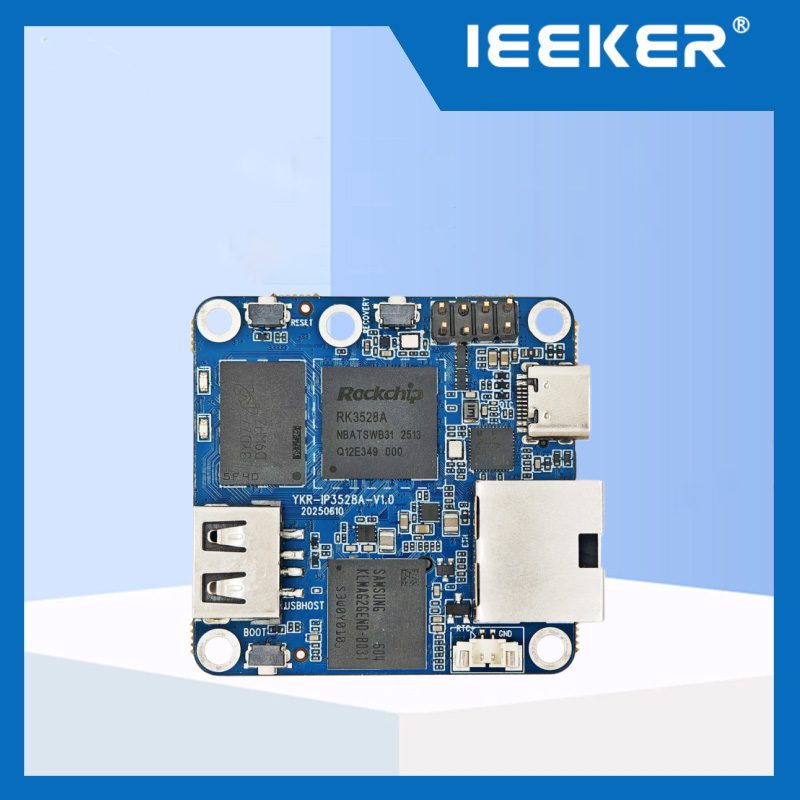
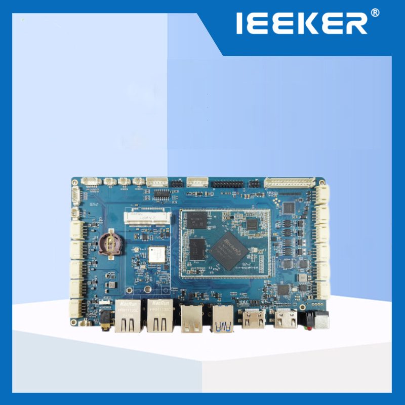
🔧 SBC industrial (placa de desarrollo)
- Fastest path to clinical prototype validation
- Full BSP support, Linux pre-validated
- Suitable for Class I devices or research use
- External isolation board needed for patient circuits
- Good for pilot and early clinical evaluation
See our Guía SoM vs SBC for a complete comparison of form factor trade-offs for production medical device programs.
Is RK3588 the Right Platform for Your Medical Imaging Device?
- ✅Portable POCUS terminal — optimal fit. ISP + NPU + H.265 encoder + battery-viable power match POCUS requirements exactly.
- ✅AI-assisted diagnostic workstation (bedside/outpatient) — strong fit. 4K display, 6 TOPS NPU, DICOM/PACS integration via Linux stack.
- ✅Portable endoscopy system — strong fit. MIPI CSI 4K input, NPU polyp detection, hardware H.265 DICOM recording.
- ✅Offline AI diagnostics (no cloud) — strong fit. On-device NPU inference eliminates PHI transmission, simplifying HIPAA/GDPR compliance.
- ✅Multi-parameter patient monitor with AI alerting — strong fit. Multiple sensor interfaces, NPU for arrhythmia models, display rendering.
- ⚠️Dermatoscopy / ophthalmology imaging — workable with ISP calibration. Requires application-specific tuning (see factory floor case study above).
- ⚠️High-volume DICOM archive (100+ GB/day) — workable with NVMe via PCIe. Plan for NVMe SSD in carrier board design; eMMC alone is insufficient.
- ❌3D MRI/CT reconstruction — not suitable. Requires GPU-class compute. RK3588 handles 2D slice viewing, not real-time volumetric reconstruction.
- ❌Radiation therapy control (SIL3+) — not suitable as sole controller. Requires dedicated safety-certified hardware separate from the application processor.
Preguntas frecuentes
Is the RK3588 IEC 60601-1 certified?
No — and it does not need to be. IEC 60601-1 certification applies to complete medical electrical equipment, not individual components or SoCs. ieeker's boards hold CE and FCC declarations as electronic components. The medical device OEM is responsible for IEC 60601-1 compliance of the finished device through proper isolation design between patient-connected circuits and the computing platform.
Can RK3588 handle real-time ultrasound beamforming?
No. Ultrasound beamforming requires dedicated FPGA or ASIC hardware with sub-nanosecond timing precision. The RK3588 operates downstream of the beamformer — receiving digitized, beam-formed image data and applying AI processing, display rendering, and DICOM archiving. A custom analog front-end board with ADCs and a beamforming FPGA sits between the probe and the RK3588.
How does on-device AI inference simplify HIPAA compliance?
When AI inference runs on-device via the RK3588 NPU, patient image data never leaves the device during processing — eliminating the cloud service provider from the compliance scope entirely. Local DICOM storage uses device-level AES-256 encryption (supported by RK3588's hardware crypto engine). Network transmission to PACS occurs only within the hospital's internal network, already within the organization's existing compliance framework.
What is the recommended OS for medical imaging devices on RK3588?
Debian 12 is recommended for devices requiring regulatory submissions. Its predictable security update cadence and apt-based package management simplify software version control documentation required for IEC 62304 compliance. Android is not recommended for Class IIa or above devices due to AOSP's complex update lifecycle. See our Guía Linux vs Android en RK3588 for the full comparison.
ieeker's RK3588 industrial SBCs and core boards provide the computing foundation for portable medical imaging devices — with pre-validated Debian/Ubuntu BSPs, DCMTK and RKNN-Toolkit2 integration support, SoM form factor options for custom carrier board designs, and engineering team availability for medical application consultation.
Fuentes y referencias
- Global Portable Ultrasound Market to Reach USD 3.84 Billion by 2030 — MarketsandMarkets (April 2026)
- Ultrasound AI Market to Reach USD 6.88 Billion by 2030 — MarketsandMarkets (2025)
- Ultrasound AI Market: 88% Clinical Integration Rate — SNS Insider (2025)
- DICOM — Digital Imaging and Communications in Medicine — Wikipedia
- HL7 FHIR — Health Level 7 Fast Healthcare Interoperability Resources — Wikipedia
- DCMTK — DICOM Toolkit, OFFIS Institute for Information Technology
- Rockchip RK3588 Brief Datasheet — rock-chips.com
- RKNN-Toolkit2 SDK — Rockchip GitHub
- IEC 60601-1 Medical Electrical Equipment Standard — IEC